Cellometer Auto 2000 Cell Viability Counter
Touchscreen fluorescent automated cell counter for primary samples
Why Auto 2000?

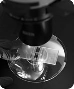
Introduction to the Cellometer Auto 2000 Cell Viability Counter
Simple, User-friendly Procedure
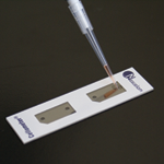
Pipette 20µl
Insert Slide
Select Assay & Click Count
View Results in 30 seconds!
Simple, Automated Cell Counting in 30 Seconds
The Auto 2000 utilizes bright field imaging and dual-fluorescence imaging to quickly and accurately identify and count individual cells. Cell count, concentration, diameter, and % viability are automatically calculated and reported.
The Auto 2000 Allows Users to:
- Increase throughput
- Increase accuracy
- Improve consistency
- Ensure all data is correctly captured
- Count difficult cells (clumpy, irregular-shaped)
- Eliminate judgment errors, miscounts, interference from red blood cells and user-to-user variability
Primary Cell Analysis: PBMCs, Stem Cells, and more
The Cellometer Auto 2000 is specifically optimized for analysis of primary cells from peripheral blood, cord blood, bone marrow, and other complex samples for use in a wide range of research areas, including:
- Nucleated Cells for Transplantation
- PBMCs for Immunology
- Splenocytes for Vaccine Development
- Stem Cells for Cellular Therapy
- Tumor Cell Suspensions for Oncology
Dual-color fluorescence allows for staining of live and dead nucleated cells, generating accurate viability results even in the presence of debris, platelets, and red blood cells. Accurate analysis of both 'messy' and 'clean' samples enables the Auto 2000 to evaluate samples at a variety of points throughout sample processing - from initial collection, to separation, to cryopreservation.
The Cellometer Auto 2000 features one-touch assays for analysis of a wide range of primary samples, including:
Nucleated Cells from Whole Blood
PBMCs / Splenocytes following Separation
Stem Cells
Tumor and Tissue Cell Suspension
No Interference from Red Blood Cells, Platelets, or Debris
The dual-fluorescence AO/PI method utilizes nuclear staining dyes that bind to nucleic acids in the cell nucleus. Because most mature mammalian red blood cells do not contain nuclei, only live and dead mononuclear cells produce a fluorescent signal. There is no need to lyse red blood cells, saving time and eliminating an extra sample preparation step. Red blood cells, platelets, and debris are not counted in the fluorescent channels.
The advantage of fluorescent counting for primary cells
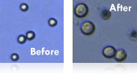
These images (right) demonstrate the advantage of fluorescent counting for primary cells. The bright field image shows the combination of nucleated cells, red blood cells, and platelets present in the sample. Only the live and dead nucleated cells are visualized and counted in the green and red fluorescent channels.| Sample | Measurement | Total nucleated | All | RBC | % RBC | n |
|---|---|---|---|---|---|---|
PBMC+RBC |
Mean |
1.26E+07 |
1.39E+07 |
1.23E+06 |
8.9% |
10 |
|
CV |
6.2% |
8.8% |
|
|
|
PBMC+1/2RBC |
Mean |
1.21E+07 |
1.27E+07 |
5.82E+05 |
4.6% |
10 |
|
CV |
4.8% |
5.4% |
|
|
|
PBMC+1/4RBC |
Mean |
1.22E+07 |
1.24E+07 |
2.27E+05 |
1.8% |
10 |
|
CV |
7.9% |
7.3% |
|
|
|
Fresh human PBMCs (peripheral blood mononuclear cells) were spiked with varying amounts of RBCs (red blood cells.) All cells (nucleated + RBC) were counted in the bright field channel. Nucleated cells were then counted in the green fluorescent channel. Varying amounts of RBCs (1.8%, 4.6%, and 8.9%) did not affect the nucleated cell count.
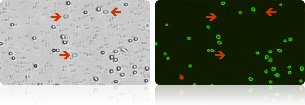
Several red blood cells are indicated in the bright field image (above, left). The red blood cells are not visible in the fluorescent image (above, right) detecting cells stained with nuclear staining dye.
Analysis of Clumpy and Irregular-shaped Cells
Including NCI-60 and clumpy MCF-7 Cells
NCI-60 is a group of 59 human cancer cell lines (originally 60) developed by the National Cancer Institute for screening purposes.
- 57% of the NCI-60 cell lines are clumpy, contain debris, or display large variations in cell shape or size
- All 59 NCI-60 cell lines have been successfully validated on the Cellometer Cell Counter
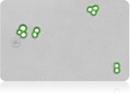
Clumpy Cells
The MCF-7 breast cancer cell line can be very clumpy. The Cellometer pattern-recognition software identifies and counts individual cells within these cell clumps for accurate analysis (shown above).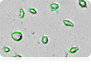
Irregular-shaped Cells
The Cellometer cell roundness setting can be adjusted for recognition and counting of irregular-shaped cells, such as RD cells and activated T-cells.User-Friendly Touch Screen
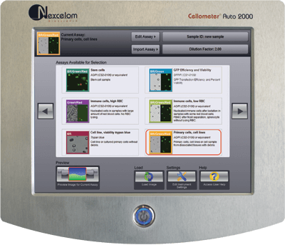
The user-friendly touch screen displays pre-optimized assays for common cell types. User-specific protocols can be easily created and saved to the menu. The simple, pre-optimized assays make the instrument easy to operate and simplify training for new users.
With the click of a button, researchers can select a new assay with saved counting parameters, making the instrument ideal for labs testing a variety of cell types. Auto-save options allow users to quickly test a series of samples.
Primary Cell Analysis: PBMCs, Stem Cells, and more
The Cellometer Auto 2000 is specifically optimized for analysis of primary cells from peripheral blood, cord blood, bone marrow, and other complex samples for use in a wide range of research areas, including:
- Nucleated Cells for Transplantation
- PBMCs for Immunology
- Splenocytes for Vaccine Development
- Stem Cells for Cellular Therapy
- Tumor Cell Suspensions for Oncology
Dual-color fluorescence allows for staining of live and dead nucleated cells, generating accurate viability results even in the presence of debris, platelets, and red blood cells. Accurate analysis of both 'messy' and 'clean' samples enables the Auto 2000 to evaluate samples at a variety of points throughout sample processing - from initial collection, to separation, to cryopreservation.
The Cellometer Auto 2000 features one-touch assays for analysis of a wide range of primary samples, including:
Nucleated Cells from Whole Blood
PBMCs / Splenocytes following Separation
Stem Cells
Tumor and Tissue Cell Suspension
Live / Dead Nucleated Cell Counts using Dual-Fluorescence
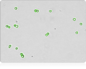
Green fluorescent live cell image

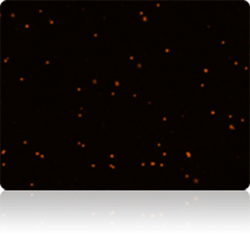
Red fluorescent dead cell image
Why Dual-Fluorescence?
Because bright field cell counting does not differentiate nucleated from non-nucleated cells and trypan blue staining is not as easy to detect as fluorescent staining, dual-color fluorescence is strongly recommended for accurate viability analysis for primary cells. The Auto 2000 is equipped with standard assays for dual-fluorescence analysis of primary cells stained with AO/PI.
The AO/PI Method
Acridine Orange, AO, is a nuclear staining (nucleic acid binding) dye permeable to both live and dead cells. It stains all nucleated cells to generate green fluorescence. Propidium iodide, PI, can only enter dead cells with compromised membranes. It stains all dead nucleated cells to generate red fluorescence. Cells stained with both AO and PI fluoresce red due to quenching, so all live nucleated cells fluoresce green and all dead nucleated cells fluoresce red.Cell Images for Data Verification
No two cells are the same.
With the Auto 2000 Cell Counter, cell morphology can be immediately viewed on-screen in the bright field image.
Counted cells are indicated on-screen for further verification that cells in the sample are being imaged and analyzed properly. Bright field counted images can be viewed for basic cell counting and trypan blue viability.
Fluorescent counted images indicating counted live and dead nucleated cells can be viewed for dual-fluorescence primary cell viability assays.
Users can confirm that:- cells are counted correctly, based on size and shape
- cells within clumps are being counted individually
- red blood cells, platelets, and debris are being excluded from results
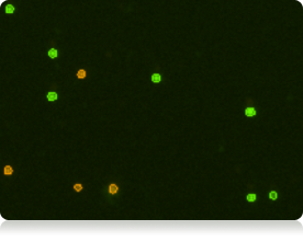
The bright field image confirms that individual cells within pairs are being counted and smaller debris is not being counted. In the combined fluorescent counted image, live counted cells are circled in green. Dead counted cells are circled in red.
- Cell images can be archived and exported for use in publications and presentations.
- Saved images can be re-counted using default or user-optimized analysis settings
Cell Size Analysis & Size-based Counting
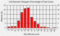
The Auto 2000 Cell Counter Automatically generates a cell size histogram based on cell diameter.
Because Cellometer generates individual cell size measurements, multiple samples can be overlaid on one histogram enabling analysis of the change in cell diameter over time.
For experiments involving SF9 or SF21 cells, saved images can be viewed and re-counted to analyze changes in morphology over time.
The minimum and maximum cell diameter settings can be optimized to count specific cells in a sample.
This example demonstrates counting of mature dendritic cells cultured from PBMCs based on cell diameter.
10x Faster than Manual Counting
Counting 1 x 10⁶ cells takes approximately 5 minutes with a manual hemacytometer. Counting live and dead cells sometimes takes twice as long. The Cellometer Auto 2000 Cell Viability Counter calculates cell count and concentration for live and dead cells and % viability in just 30 seconds.
Improve Data Accuracy & Consistency
- Eliminate Wash Steps
- Eliminate Judgment Errors
- Eliminate interference from RBCs
- Eliminate Recording & Calculation Errors
- Reduce Counting Time … Run More Experiments
Cellometer Precision
The Cellometer Auto 2000 offers excellent reproducibility, with a %CV (Coefficient of Variation) of <10% for fluorescent concentration and viability measurements. The data (right) is based on four preparations of Jurkat cells stained with propidium iodide, a fluorescent nuclear-staining dye.Sample |
N Value |
Average Live Cell Concentration |
% Viability |
CV of Concentration |
CV of Viability |
|---|---|---|---|---|---|
A |
4 |
4.20E+06 |
91.1 |
10% |
2% |
B |
4 |
1.06E+06 |
22.7 |
7% |
1% |
C |
4 |
3.27E+06 |
57.5 |
7% |
7% |
Imaging / Counting Chambers: No Washing or Contamination
The disposable Cellometer Cell Counting Chambers offer several key advantages:
- Time savings - no washing
- No risk of cross-contamination
- Reduced biohazard risk to users
- Controlled sample volume
- Large-depth chambers for large cells
- Most affordable automated counting consumables
Auto-Save Data and Cell Images
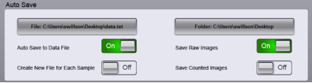
The Auto 2000 allows users to generate specific folders to which data reports and images can be auto-saved. Using the auto-save feature ensures all data is accurately captured in the correct location. It also enables users to test a series of samples very quickly.
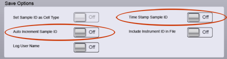
Users can configure the Auto 2000 to request a new sample name for each sample that is imaged and counted or apply auto-numbering to individual samples in a series to further reduce analysis time. Users can also apply a time stamp to individual data files.
Dedicated On-line and On-site Applications Support
- Creation of new cell types
- Optimization of counting parameters
- Troubleshooting
- Training of new users
- Installation of the Auto 2000 Cell Viability Counter

The help button at the bottom right of the Cellometer Auto 2000 software screen gives users instant access to:
- Software features and instructions
- On-line tutorials and training videos
- Submission of a Support Ticket
All Nexcelom Applications Specialists are 100% focused on image-based cell concentration & viability and cell-based assays using Cellometer Image Cytometry.
Nexcelom Field-based Applications Specialists are also available for:
- On-site demonstrations
- Training
- Troubleshooting
- Technical Seminars